1.5 Step 10. Reflection of the tentorium cerebelli and removal of the brain
Hellow guys, Welcome to Utubetrends, and you are watching 1.5 Step 10. Reflection of the tentorium cerebelli and removal of the brain. and this vIdeo is uploaded by Netter's Anatomy Dissections at 2017-10-20T06:55:36-07:00. We are pramote this video only for entertainment and educational perpose only. So, I hop you like our website.
Info About This Video
| Name |
1.5 Step 10. Reflection of the tentorium cerebelli and removal of the brain |
| Video Uploader |
Video From Netter's Anatomy Dissections |
| Upload Date |
This Video Uploaded At 20-10-2017 13:55:36 |
| Video Discription |
These videos have been excerpted from Netter’s Video Dissection Modules on Student Consult. http://bit.ly/2oEt9CO
Step 10
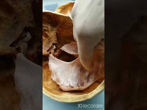
With the falx cerebri peeled posteriorly, note where the great cerebral vein of Galen joins the straight sinus. This connection was torn when the dura was posteriorly reflected. The falx cerebri should remain attached posteriorly to the tentorium cerebelli, which can now be reflected as a result of the previous cuts. With the vertebral arteries and all of the cranial nerves transected, and the spinal cord cut in an earlier dissection, the brain can be gently removed from the skull. Place the brain in a bucket of fixative to preserve it.
Key Terms
• Falx cerebri: a vertical, midline extension of the dura mater N104 N110. It lies between the cerebral hemispheres and fuses with the tentorium cerebelli posteriorly and attaches to the crista galli anteriorly. The superior sagittal sinus is located along its superior boundary with the dura mater and the inferior sagittal sinus courses within its inferior margin. The straight sinus occurs along its border with the tentorium cerebelli.
• Great cerebral vein (of Galen): lies in the midline, dorsal to the mesencephalon and between the posterior margins of the corpus callosum and cerebellum. This vein penetrates the posterior margin of the tentorial incisure to join the straight sinus N145 N146. This vein, with its connection to a fixed dural substrate, is most susceptible to tears in infants during head manipulation at birth or when the infant is shaken. The main internal cerebral veins are tributaries of the great cerebral vein.
• Straight sinus: the dural sinus that begins at the center of the anterior margin of the tentorium cerebelli and extends straight posteriorly along the line of fusion of the falx cerebri with the tentorium cerebelli N104. Anteriorly, it receives the great cerebral vein of Galen and the inferior sagittal sinus, while posteriorly, it contributes to the confluence of sinuses N105. Superior cerebellar veins drain into its inferior surface.
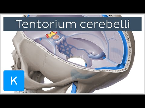
• Tentorium cerebelli: the partition formed of dura mater that forms the roof of the posterior cranial fossa N105. It separates the cerebellum from the cerebral hemispheres and is fused with the posterior inferior limit of the falx cerebri. The straight sinus occurs along the line of fusion N146. The tentorium is attached to the clinoid processes, along the crests of the petrous ridges, along the grooves for the transverse sinuses, and to the internal occipital protuberance. It has a large opening, the tentorial incisure, for passage of the brainstem. One of the more important relationships is the passage of the oculomotor nerve over the free edge of the tentorium near the posterior clinoid process. Here the nerve can be compressed by the brain when there is an increase in intracranial pressure that forces the brain into the incisure (tentorial herniation).
• Vertebral artery: a branch of the subclavian artery on either side of the body. It passes superiorly through the transverse foramen (foramen transversarium) of each cervical vertebra except C7, until it reaches C1 N22 N23. Here the artery travels across the posterior arch of C1 N175 before entering the foramen magnum. Inside the skull, the two vertebral arteries join up to form the basilar artery N167 N141 N142 at the base of the medulla oblongata. The basilar artery is the main blood supply to the brainstem.
• ABOUT: The project was made possible by several very dedicated faculty and staff at University of North Carolina, Chapel Hill--especially O.W. Henson and Noelle A. Granger--and partner schools, and by a grant from the Fund for the Improvement of Post-Secondary Education of the US Department of Education. This channel includes over 400 short videos highlighting the steps in a full-body human dissection in the gross anatomy lab. Each step is narrated and key structures labeled. |
| Category |
People & Blogs |
| Tags |
People & Blogs Download MP4 | People & Blogs Download MP3 | People & Blogs Download MP4 360p | People & Blogs Download MP4 480p | People & Blogs Download MP4 720p | People & Blogs Download MP4 1080p |
More Videos